

Osteoporosis is often called a silent disease because it develops gradually and usually produces no symptoms until a fracture occurs. The first indication may be a low-impact fracture, noticeable height loss, or curvature of the spine (kyphosis). In advanced stages, osteoporosis-related bone thinning can sometimes be detected on routine X-rays.
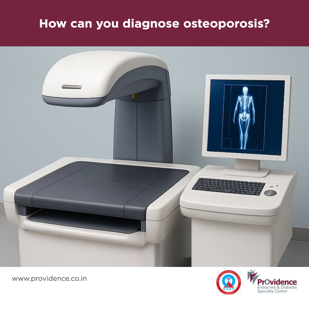
For screening purposes, peripheral dual-energy X-ray absorptiometry (DXA) is frequently used at health camps. This quick and non-invasive test measures bone density at the heel and can help identify individuals who require further evaluation. If the results are suggestive of bone loss, a central DXA scan is recommended. This test uses a small, safe dose of X-rays to measure bone mineral density (BMD) at key fracture-prone sites—the spine, hip, and forearm. The BMD result is expressed as a T-score, which compares an individual’s bone density to that of a healthy young adult. The T-score helps classify bone health as normal, osteopenic, or osteoporotic.
In some research or specialized clinical settings, quantitative computed tomography (QCT) may be used to assess bone density in three dimensions, though this is less common in routine clinical practice.
Dr. Mathew John, MD, DM